|
Department of Pathology and Cell Regulation (PCR) Kyoto Prefectural University of Medicine |

|
-Research-
Biomedical optics and photonics for molecular and pathological analysis techniques
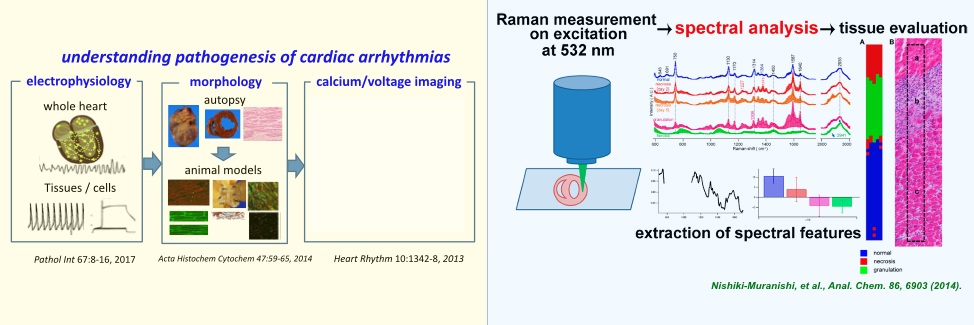
Our department has kept learning advanced optics and photonics technologies to lead interdisciplinary research of medicine and optics/photonics. Our recent interests are Raman spectroscopy for label-free and minimally invasive, pathological evaluation of ischemic heart disease and fatty liver disease, and peripheral nerve detection against its adjacent tissues in surgery, 5-aminolevlinic acid for gastrointestinal primary and metastatic cancer detection, by protoporphyrin IX fluorescence, and UV imaging for specific cancer detection and nucleic acid detection. Reference S. Ohira, et al., Sci. Rep. 7, 42401 (2017). Y. Kumamoto, et al., Sci. Rep. 7. 845 (2017). N. Koizumi, et al., Eur. J. Surg. Oncol. 42, 1236 (2016). Y. Kumamoto, et al., Biomed. Opt. Express 7, 158-170 (2016). T. Minamikawa, et al., Sci. Rep. 5, 1716 (2015). N. Nishiki-Muranishi, et al., Anal. Chem. 86, 6903-6910 (2014). T. Minamikawa, Histochem. Cell Biol. 139, 181-193 (2013). K. Imaizumi, et al., Gastrointest. Endosc. 75, 110-117 (2012). Y. Harada, et al., Histochem. Cell Biol. 132, 39-46 (2009). M. Ogawa, et al., Biochem. Biophys. Res. Commun. 382, 370-374 (2009). Y. Murayama, et al., Int. J. Cancer 125, 2256-2263 (2009). Visualization of dynamics of functional molecules in the heart The overall objective of our research is to better understand the pathophysiology of the heart especially arrhythmias via visualization of functioning molecules in the cardiomyocytes. Using our rapid-scanning scanning confocal technologies, we have been engaging mainly in visualization of intracellular calcium signals in the heart with high spatiotemporal resolutions. Of various functioning molecules, calcium ions play pivotal roles in excitation and contraction of the heart. In the intact heart the individual cardiomyocytes exhibit intracellular calcium transients (transient rise of intracellular calcium concentration) uniformly on electrical excitation, whereas under pathological conditions heterogeneous calcium dynamics arise among individual myocytes, e.g., wave-like propagations of the intracellular calcium in the cells (calcium waves) and beat-to-beat alterations of calcium transients (calcium alternans). These abnormal calcium dynamics, originated from altered functions of various calcium-handling proteins (e.g., ryanodine receptors, calcium pumps, and sodium-calcium exchanger) are responsible for abnormal excitation/conduction and contraction leading to arrhythmias and contractile failure. Moreover, intracellular calcium ions have additional impact on cell-cell communication via gap junctions, abnormality of which also contributes to generation of arrhythmias. Current on-going integrated researches involve ultra-rapid confocal visualization of membrane potentials and calcium in addition to immunohistochemical imaging of various functioning proteins that regulate cell-cellular and cell-extracellular milieus. Reference H. Tanaka, et al., Pathol. Int. 67, 8-16 (2017). Y. Jiang, et al., Acta Histochem. Cytochem. 47, 59-65 (2014). T. Matsuyama, et al., Heart Rhythm 10, 1342-1348 (2013). K. Fujiwara, et al., Circ. Res. 103, 509-518 (2008). T. Nakagami, et al., Cardiovasc. Res. 79, 70-79 (2008). T. Hamamoto, et al., J Mol. Cell Cardiol. 38, 561-569 (2004). H. Tanaka, et al., J. Mol. Cell Cardiol. 34, 1501-1512 (2002). T. Kaneko, et al., Circ. Res. 86, 1093-1099 (2000). |